An Introduction to the Laboratory
Microscopy
The microscope is perhaps the most basic tool used by biologists. Light microscopy
was first applied to biological materials in the seventeenth century by such
researchers as Robert Hooke and Antoni van Leeuwenhoek. The impact of this invention
on the course of human endeavor is easily underestimated. Within a short span
of time, that which was invisible to the human eye became visible. Events such
as the causes of certain diseases and the decay of food, previously described
as spontaneous generation or supernatural could now be explained. An entire
world of living organisms previously unknown was now available for study. The
microscope remains a pivotal tool in learning biology to this day, with most
laboratories being equipped with one or more research instruments.
Over the course of this year, you will be using two different types
of microscope:
 OR
OR 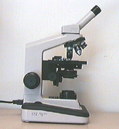
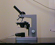
These microscopes are delicate and expensive instruments and must be treated
with care.
When using a Compound Microscope please keep the
following in mind:
- When transporting your microscope always pick it up by the arm. Hold
it upright and support the base with your free hand.
- The arm supports an inclined body tube, which in turn supports the
magnifying elements of the microscope. At the end of the tube is the ocular,
one of the magnifying elements. The other magnifying elements are screwed
into a revolving nosepiece at the bottom of the body tube; these are called
objectives.
Together, the ocular and an objective constitute the magnifying system of
the microscope. The objective provides for the primary or initial magnification
of the object being viewed while the ocular provides the secondary or final
magnification and generates the image seen by the viewer.
- Underneath the body tube and nosepiece is a flat plate, the stage,
upon which objects to be examined are placed. Directly beneath the stage opening
you will notice a system of lenses, the condenser,
which serves to concentrate the light from the illuminator
below.
- Light plays an extremely important role in the operation of a compound microscope.
The critical importance of light makes necessary its careful adjustment, and
several controls are available for this purpose. The iris diaphragm may
be found just below the condenser lens system. This control is one of the
most important on your instrument.
- Just as with a magnifying glass, viewing an object with the microscope requires
that the lens be a certain specified distance from the object. That distance
is a property of the lens system, and is constant for any particular objective;
it is called the working distance. At the working distance
from an object, the objective is in focus. Changes and adjustments in the
focus, which are necessary when an object is first placed on the microscope
and essential in order to perceive depth, are accomplished by means of coarse
and fine adjustment
knobs usually located on the arm. Note that the fine and coarse focus knobs
are separate on some of our microscopes and combined on others.
You should be able to locate the terms in bold on the image of the light
microscope above, or click
here if you are having problems.
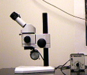
Keep the following in mind when using a Stereoscopic Microscope:
- The depth of field is much greater than with the compound microscope, which
allows objects to be view in three dimensions
- The optics do not invert images
- To focus close your left eye and adjust the focus of the image viewed
with the right eye. Open your left eye and close your right eye and adjust
the focus for the left eye using the secondary focus ring located on
the left ocular.
- All of our stereo microscopes are binocular. To obtain the correct
interpupilary distance for your eyes you must adjust the distance between the
two oculars by gently pushing or pulling the oculars until you see the object
as a single image.
- Note that the dissecting microscope is not parfocal and must be re-focused
when the magnification is changed
Consider the following questions for both the light microscope and the
stereoscopic microscope
- When you move the slide to the right while looking into the microscope, which way does the image appear to move?
- Which way does the image move when you move the slide toward you?
- Away from you?
Click here to view the letter F slide as seen through a compound light microscope
Click here to view the letter F slide as seen through a stereoscopic microscope
First published Sept 95: Modified June 2019
Copyright © Michael Shaw 2019 (Images and Text)
 Return
to Biology Home
Return
to Biology Home  Back
to Lab Index
Back
to Lab Index  Forward
to Next Page
Forward
to Next Page  University
of Manitoba Home
University
of Manitoba Home