 |
Phylum Platyhelminthes
(Canadian Campbell 2nd ed Pages 734 - 736)
 |
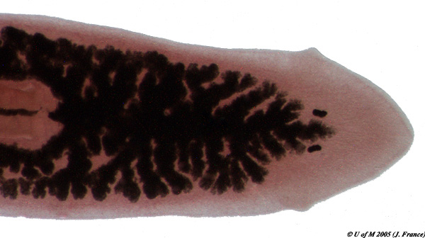
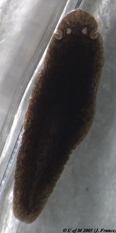
This phylum contains three classes, the Turbellaria which are free-living flatworms, the Trematoda or flukes and the Cestoda or tapeworms. The Trematoda and Cestoda are all parasitic in their mode of existence. All forms are bilaterally symmetrical (image) and dorsoventrally flattened. Between the ectoderm and the endoderm surrounding the enteron (gastrovascular cavity) lies a loose tissue, the mesoderm, which represents an advance over the Cnidarians. Flame cells are present in the excretory system. Platyhelminthes lack a body cavity and are thus referred to as acoelomate (image). Both male and female sexual organs are frequently found in one individual (hermaphrodite). Examine a whole mount of a planarian External features to note are: head region, eyespots, projecting auricles and ventral proboscis. |
Consider the following:
The planarian that you will be studying in this lab belongs to the genus Dugesia.
Examine
the whole mount of Dugesia
Examine
the anterior end of Dugesia at 10x
Observe the enteron (gastrovascular cavity). It has one anterior and two posterior caeca with numerous side branches (diverticula). The proboscis can be seen in its retracted position in the proboscis chamber. The pharynx lies within the proboscis and connects to the enteron. The mouth is at the end of the proboscis.

Reproduction is by both sexual and asexual means. For sexual reproduction, planarians are hermaphroditic organisms.
Asexual reproduction is accomplished by fragmentation followed by
regeneration, and many of these free-living flatworms have remarkable
regenerative abilities. As a result of this, the turbellarian flatworms have
contributed a great deal of information to our knowledge of regeneration.
Most of the species used in these studies are members of the family
Planariidae and are thus generally known as planarians.
The regenerative capacities of planarians have long been recognized, and the
studies are of particular significance because these animals display bilateral
symmetry, and organ level of organization and their method of regeneration
is in some respects similar to regeneration in vertebrate animals. Regeneration
experiments in planarians have shed light on such interesting processes as memory
and learning.
Regeneration of a new individual from the bisected parts of a healthy planarian
normally takes about two weeks or more depending on the temperature. Regeneration
is preceded by the formation of a blastema
which is recognized as a small blob of clear cells at the cut surface. The blastema
is composed of relatively undifferentiated cells which presumably arise from
special cells in the parenchyma,
called neoblasts.
Neoblasts are RNA rich cells which are undifferentiated and apparently set aside
early in the embryology of the planarian. The blastema and newly generated parts
of planarians are generally characterized by lack of pigmentation and are thus
readily distinguished from the older body parts.
Regeneration in planarians takes place under natural conditions as well as artificially in the laboratory. Under natural conditions the planarian ordinarily fragments into two parts, with the line of fission occuring just posterior to the pharynx. The anterior part then regenerates a new tail while the posterior fragment develops a new head. Therefore, this is a form of asexual reproduction. Recent studies have shown that natural fragmentation occurs more frequently when populations of planarians are low and less frequently under crowded conditions.
Consider the following:
Examine a prepared C.S. of Dugesia through a region of the body showing the pharynx.
The outside cellular layer of the surface of the body is derived from ectoderm, and consists chiefly of cuboidal epithelial cells. These cells form the epidermis. Find the layer of circular muscles just beneath the epidermis. Immediately below the layer of circular muscles are the longitudinal muscles which have been cut transversely and appear as tiny irregularly shaped masses of tissue. These are derived from the mesoderm. Observe sections of the diverticula and of the enteron; each can be identified as a cavity lined mainly with tall, columnar epithelial cells with large vacuoles. This is the endoderm. Filling the space between the other tissues is a peculiar connective tissue known as parenchyma which is derived from mesoderm.
The flukes are all parasitic organisms; the adult stage living in or on a wide variety of vertebrate animals. In general, flukes tend to have rather complex life cycles involving two or more hosts. Of all the groups, the blood flukes are the most important as they are the causative agents for the human disease known as Schistosomiasis. This debilitating disease affects some 400,000,000 persons in Asia, Africa, parts of South America and the Caribbean (Campbell 6th Ed. 653; 7th Ed. 647).
Another important fluke is Fasciola hepatica, a bile duct parasite of sheep and cattle.
Examine a preserved specimen of the sheep liver fluke Fasciola hepatica
Tapeworms are elongate, intestinal parasites consisting of an anterior scolex for attachment and numerous repeating body segments called proglottids. They are highly modified to a parasitic existence and have no digestive system. When mature, each proglottid contains thousands of eggs. They are released from the posterior end of the tapeworm, leaving the host's body with feces. Tapeworms also have complicated life histories.
Examine
a prepared slide of a tapeworm scolex
(Note the hooks and suckers for attachment to the host)
Examine
preserved tapeworm proglottids
(Note that each segment of the tapeworm body is an entire reproductive unit)
![]()
