 |
The Rat
(A Representative Chordate and Mammal)
|
Two common species of the genus Rattus, the Black Rat (R. rattus) and the Brown or Navy Rat (R. norvegicus), are similar in size and appearance, and therefore, difficult to tell apart by superficial examination. This problem is even greater in many laboratory specimens, since mutant white albinos from either species are commonly used. As is often the case, however, closer examination of one or more characteristic external features, even in albinos, provides sufficient information to permit species identification. In Rattus spp. the ratio of tail length to body length differs between species. In R. rattus, the tail length is typically greater than the body length; while in R. norvegicus, the tail length is typically less than the body length.
A typical chordate has a number of distinctive characteristics, both external and internal. In this portion of the laboratory, we shall be concerned with the former.
The chordates are bilaterally symmetrical, show a high degree of cephalization with a close association between the brain and special senses, have a definite trunk region with associated appendages, have an anterior mouth and a post-anal tail. Most of these characteristics reflect the active mode of life pursued by the chordates, in which locomotion is typically directed anteriorly, parallel to the longitudinal body axis. However, modifications to this plan are numerous and in many cases there is a strong link between the degree of modification and the animal's environment.
The rat is a typical mammal, with head, clearly defined neck, trunk and tail; the trunk is further divided into the anterior thorax (chest region, containing the heart and lungs) and the posterior abdomen. There is a terminal mouth with lips, and external nares immediately above. The ear is superficially (see definition #16) represented by the exernal ear consisting of the outer, flexible, cartilaginous pinna, and a short canal, the external auditory meatus, leading to the internally situated ear.
As a typical mammal, the rat posses two unique external characteristics that distinguish mammals from all other vertebrates:
The rat, like many other mammals possesses special stiff sensory hairs, the vibrissae,
on the sides of the face.
Click
here to examine the vibrissae of Rattus spp.
Mammary glands are present in both sexes, but may be difficult to locate on the male and immature specimens. Typically three pairs of teats will be found on the ventral thoracic region, and an additional three pair posteriorly, on the ventral abdominal surface. The limbs of the rat are pentadactyl and tripartite; the hind limbs larger than the fore limbs. Note the terminal claws. The rat's gate, or mode of locomotion is called digitigrade, because the animal walks only on the digits of the foot, the remainder being held off the substrate. (In contrast, except for minor areas such as the instep, the entire foot of the human is placed on the substrate during walking, hence the gait is called plantigrade). Note the scales on the tail.
Examine
the female rat externally
Examine
the male rat externally
Before examining the skeletal muscles of the rat, carefully examine the skeletons of the following:
Pay particular attention to the shape of the cranium (head), the vertebral column, pectoral and pelvic girdle , and the fore and hind limbs. Observe closely the similarities in the skeletal structures between the four classes, Pisces (fish), Amphibia (frog), Aves (bird) and Mammalia (rat). List some similarities. Also note some differences in the shape of the pectoral (anterior) and pelvic (posterior) girdles and position of limbs relative to the girdles. You should be able to identify the bones of the fore limb (humerus, ulna and radius) and the bones of the hind limb (femur, tibia and fibula). Differentiate between cervical (neck region), thoracic (rib region) and lumbar (abdominal region) vertebrae.
Many people are not aware that bone is a living tissue. Bone contains living cells and has a nervous supply along with a vascular supply. Bone can be added to or depleted. In fact bone tends to decalcify with age and become brittle. This is especially true in the human female. Diet and exercise will help to counteract this process. Bone is a tissue in which the extracellular component is highly calcified. It is close to cast iron in tensil strength but is less than one third the weight.
Examine
the bone cross section x10
Examine
the bone cross section x40
On viewing you will notice a number of relatively large dark ovals or circles with a concentric arrangement of tissue around the outside. The central dark oval or circle is known as a Haversian canal. The Haversian canal contains elements of the vascular system to provide nutrients to the bone and remove waste materials from the bone. The concentric layers around the canals are called lamellae. Uniformly spaced along the lamellae are small cavities called lacunae. Each lacuna appears as a dark oval in these preparations but in living bone each contains one bone cell known as an osteocyte. Osteocytes are responsible for secreting the extracellular matrix which makes up most of the mass of the tissue. Radiating from the lacunae are very fine passages called canaliculi. In these preparations the canaliculi appear a dark thread-like lines extending out from the lacunae. There are connections between the haversian canals, the lacunae and the canaliculi. Bone is thus not solid but filled with a fine interconnecting system of canals. One haversian canal with its surrounding lamellae makes up a cylindrical haversian system.
Make a sketch of bone tissue labelling the various parts.
Muscular
System
(Canadian Campbell 2nd ed. Fig. 40.5)
Muscle tissue may be divided into three types:
We will be concerned only with some of the more easily identified, superficial skeletal muscles.

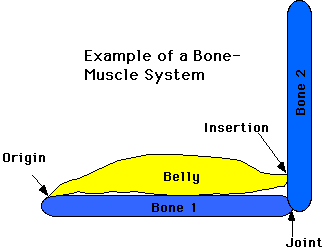
Generally, a muscle is attached at each end. The less movable attachment is called the origin, the more movable attachment, the insertion. The fleshy central portion of a muscle is called the belly. The attachment may be to a bone by means of a narrow band of connective tissue called an aponeurosis. Most skeletal muscles move bones and cartilages, some also cause movement of soft parts, for example, facial muscles which originate on a bone and insert on the easily movable skin of the face.
As you examine the images of the dissected rat identify the muscles from their descriptions below and note especially the type of movement caused when each muscle contracts. It is these movements that make possible the wide range of behaviour necessary to the survival of the living animal. Muscles are covered by and separated from each other by thin layers of connective tissue called fascia.
Muscles that cause particular types of action can be described as follows:
Extensors - straighten members such as fingers, arms, etc.
Flexors - bend members such as fingers, arms, etc.
Rotators - turn members on their axis (e.g. turn your neck sideways)
Elevators - lift or raise parts or structures
Depressors - lower or depress parts or structures
Sphincters - surround openings which close when muscles contract
Dilators - expand openings
At the posterior angle of the jaw, locate the large masseter muscle, which elevates the jaw. Note the various neck muscles that elevate, depress and rotate the head. On the chest, notice the pair of flat, triangular pectoralis muscles, one on each side of the midline.The pectoralis major originates on the sternum and inserts on the humerus. The anterior margin of each pectoral muscle is marked by the disappearance beneath it of the external jugular vein, and by the lateral passage of a superficial vein from the shoulder.
The lateral border of the pectoral muscle is somewhat disguised in the region of the armpit by the cutaneous maximus muscle which originates in the region of the armpit and the outer surface of the latissimus dorsi muscle. The cutaneous maximus muscle inserts on the skin (image). This muscle is used for shaking the skin. See how the pectoralis maximus muscle attaches to the upper arm. Its chief action is to pull the arm ventrally and to rotate it somewhat.
Originating around the shoulder joint i.e. scapula (shoulder blade) and appearing from beneath the distal attachment or insertion of the pectoralis muscle is the biceps muscle. This muscle inserts on the radius just distal to the humerus. It flexes and rotates the forearm. On the back side of the arm, antagonistic to the biceps, is the triceps muscle. It originates from the humerus and the scapula and inserts on the ulna. The triceps muscle extends the forearm. Running back from the armpit region on the medial side of the humerus, and somewhat hidden by the cutaneous maximus, is the latissimus dorsi muscle which passes posterior and dorsal. The latissimus dorsi originates on the thoracic and lumbar vertebrae and inserts on the shaft of the humerus. Its action is to pull the arm backward and upward. For each of these muscles locate the origin and insertion on the rat skeleton.
On the forearm (distal to the humerus), note the fleshy muscles near the elbow which extend as long tendons over the wrist and attach to the digits. Most muscular control of the digits comes from these muscles. Wiggle your fingers and clench your fist and watch the play of muscles in your upper forearm. You can see and feel the tendons on the front of your wrist and the back of your hand. Muscles that move your fingers are mainly located proximal to the wrist, and are connected to the digits by long tendons.
The abdominal body wall is very thin and its covering fascia is quite tough. In the mid-ventral line find a narrow, longitudinal, whitish band. This is the tendinous linea alba. On each side of it lies a longitudinal strap-like muscle, the rectus abdominis. The rectus abdominis originates on the pelvic girdle (pubis) and inserts on the cartilage of the first and second ribs. The overlying connective tissue must be cleared to see the longitudinal direction of its fibers. Lateral to the rectus abdominis are three layers of muscle, extending over the rest of the ventral and lateral portions of the abdomen. From outside to inside, these are:
![]()
