|
In the rat, as in all tetrapods, there is a sharing of the anterior structures of the digestive and respiratory system. A special gate-like structure, the epiglottis, is present at the entrance to the respiratory structures leading to the lungs, which makes possible a time-sharing arrangement between the two systems. Usually much of the oral cavity and the nasopharyngeal region perform a respiratory function, passing air in and out of the lungs, past the raised epiglottis. When food is taken into the mouth the oral cavity becomes, functionally, a part of the digestive system. The act of swallowing, resulting in the passage of food back through the pharynx to the esophagus, simultaneously causes the epiglottis to close off the respiratory system, thereby ensuring that food does not enter the passages leading to the lungs.
The vertebrate digestive system consists of the alimentary canal and its associated glands. The alimentary canal is essentially a tube running from the mouth to the anus. Specialization in both the gross anatomy and the fine structure is found along the tube to cope with the demands of the animal's food habits and with the various functions performed, (ingestion, digestion, absorption and elimination).
Pry the jaws apart to view the buccal cavity. Upon doing so, you will notice that the roof of the mouth is divided into a posterior soft palate and a bony secondary (hard) palate anteriorly. The buccal cavity contains the long, narrow, bony jaw, with teeth set in sockets in the jaw bones. The teeth of the rat, like those of other mammals, are heterodont (i.e. teeth are not all alike). Primitively, mammals teeth consist of incisors, canines, premolars and molars.
The dental formula for the rat is as follows:
1 |
0 |
0 |
3 |
1 |
0 |
0 |
3 |
|
x |
2 |
= |
16 |
The upper row refers to the upper jaw, while the lower row refers to the lower jaw. The first column corresponds to incissors; the second, canines; the third, premolars and the forth, molars. Thus, the rat has 1 incisor, 0 canines, 0 premolars and 3 molars in both the upper and lower jaw. Note that this represents only one half of the jaw. In the rat, as in other rodents, a gap called the diastema is present between the incisors and molars. Note the strikingly different shapes of the incissors and molars of the rat.
In the body cavity examine the membranous mesenteries and omenta. A mesentery is best observed attaching the small intestine to the dorsal body wall. Within this mesentery, note the blood vessels which run through it. An omentum may be observed running from the greater curvature of the stomach to the spleen. The parietal peritonium may be seen as a thin layer of connective tissue lining the abdominal cavity, while the visceral peritonium is similar tissue covering the viscera.
Examine an example of a mesentery and omentum
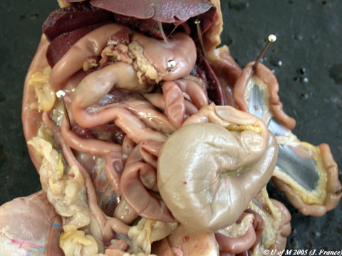
Locate the diaphragm between the abdominal and thoracic cavities. Directly posterior, note the large liver with its five lobes; the right and left central lobes to the right and left of the midline, the right and left lateral lobes, lateral to the central lobes. The left lobe is large and overlaps the stomach, while the right is a double lobe overlapping the right kidney. The rat has no gall bladder. The stomach, which is divided into the anterior cardiac and more posterior pyloric portion, lies in the left side of the abdominal cavity just below the liver. The esophagus, which runs through the thorax from the pharynx, enters the cardiac portion of the stomach. The pyloric sphincter muscle controls movement of food from the pyloric portion of the stomach to the small intestine.
Examine the rat abdominal cavity freshly opened
|
|
The duodenum is in the form of a loop with descending, transverse and ascending limbs. The pancreas and pancreatic ducts are within the bend of the duodenum. The jejunum, which is thick-walled, begins where the duodenum turns posteriorly and terminates with the commencement of the thin-walled and hence, darker colored ileum. The ileum runs to the caecum. The large intestine consists of the caecum, colon, and rectum. The rectum is that portion of the large intestine which continues through the pelvic muscles. The alimentary canal terminates with the anus.
Examine
the first portion of the alimentary canal
Examine
the large intestine of the rat
The anatomy of the digestive tract is variable depending upon the food habits of the animal. Carnivorous animals have a relatively short digestive tract. Herbivores, on the other hand, which take in large quantities of difficult to digest cellulose, have long digestive tracts, often equipped with various internal pouches or expansions where intestinal microorganisms can aid in cellulose breakdown. Large herbivores like cattle, sheep and deer have a four-pouched stomach. One of these pouches, the rumin, contains large populations of microorganisms. Food is repeatedly regurgitated from the rumen and rechewed as a cud before passing through the rest of the gut. Rodents like the rat and lagomorphs like the rabbit possess a large caecum, a blind sac which attaches to the gut at the junction of small and large intestines. The caecum is also important in cellulose digestion.
![]()