
|
Histological Material
Study the prepared slide of a cross section through the mammalian spinal cord in the region of a dorsal root ganglion.
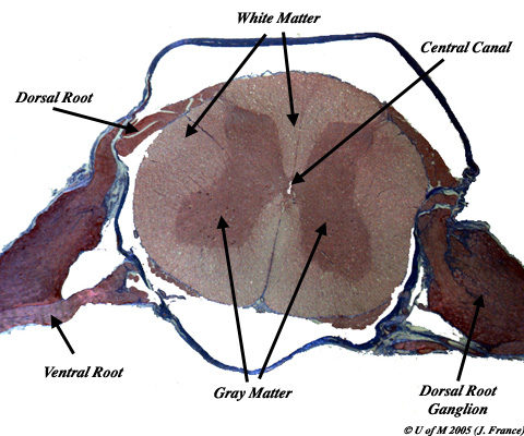
In the center of the cord locate a small, clear space which is known as the central canal. This canal is filled with cerebrospinal fluid and runs the length of the spinal cord. At its anterior end it is continuous with the fourth ventricle of the brain. In viewing the cross section you should note a butterfly shaped area of gray matter filling much of the central area. The gray matter is named because it appears gray in fresh tissue due to the presence of numerous cell bodies. The peripheral area of the section is made up of the more uniformly colored white matter. In the cross section, the white matter has a granular appearance. This is because the white matter consists largely of axons which communicate up and down the spinal cord. They appear as small, round dots in transverse section. In your section, you should also see parts of dorsal and ventral roots as well as the dorsal root ganglion which will help you to orient the section.
In the ventral region of the gray matter, notice the large nerve cell bodies. These are motor cell bodies, from which axons extend via the ventral root to muscles. Smaller cell bodies may be found more dorsally in the gray matter. These cell bodies belong to association neurons, which take impulses from incoming sensory fibers and switch them either directly to a motor neuron (in the simplest case) or to other fibers which communicate up, down or across the spinal cord.
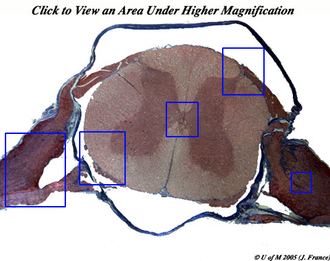
Use the Image below to zero in on the various regions:
Examine a prepared slide of isolated nerve fibers:
This slide contains a short section of myelinated axons.
![]()