
|
The Vascular Level of Organization
 The vascular plants are land dwelling plants that have been traditionally
classified into one group, the Tracheophyta suggesting a close evolutionary
relationship among the major groups of vascular plants. The vascular plants can
be artificially divided into two major groups, the seedless vascular plants
and the seed plants. The evolutionary history of the vascular plants remains
controversial with some authorities maintaining that the major groups of vascular
plants have evolved along independent lines. The classification system used in
Campbell reflects the latter view and divides the living vascular plants into
seven phyla (Canadian Campbell 2nd ed. Concept 29.3).
The vascular plants are land dwelling plants that have been traditionally
classified into one group, the Tracheophyta suggesting a close evolutionary
relationship among the major groups of vascular plants. The vascular plants can
be artificially divided into two major groups, the seedless vascular plants
and the seed plants. The evolutionary history of the vascular plants remains
controversial with some authorities maintaining that the major groups of vascular
plants have evolved along independent lines. The classification system used in
Campbell reflects the latter view and divides the living vascular plants into
seven phyla (Canadian Campbell 2nd ed. Concept 29.3).
The life cycle of a vascular plant is conceptually the same as for
bryophytes. The major difference is that the sporophyte generation
is the dominant and persistent phase of the life cycle, while the gametophyte
is reduced and short-lived.
The sporophyte
phase consists of a complex plant body composed of roots, stems and leaves.
These plant organs possess highly differentiated vascular tissues called xylem and phloem.
The growth, structure and function of these organs and tissues will be examined
in laboratories 9 and
10. The gametophytes are structurally simple, ranging from the free
living macroscopic thalli of the ferns to the
microscopic gametophytes of the seed plants.
The
Seedless Vascular Plants
Canadian Campbell 2nd ed. Concept 29.3
Structure
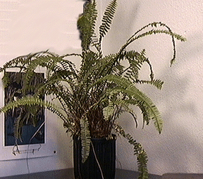
The ferns compose a group of several thousand species growing in many diverse habitats, the most common being shady places with abundant moisture. The most conspicuous part of the fern sporophyte is the leaf. The typical fern leaf, often termed a frond, is an elaborate structure composed of numerous leaflets. The frond arises from the stem which is a rhizome growing on or just beneath the soil surface. Young fronds are tightly coiled and are commonly referred to as "Fiddleheads". The rhizome also bears numerous roots, termed adventitious roots since they arise from stem tissue. This distinguishes them from the primary root (derived from the embryo) and secondary or lateral roots (derived from a pre-existing root). All tissues of the shoot (leaf and stem) are derived from a single meristematic cell located at the apex of the rhizome. Similarly all tissues of the root are derived from a single meristematic cell located at the apex of the root. The fronds, the rhizomes and the roots make up the sporophyte generation.
Examine a fern sporophyte and note the fronds
When mature, the leaflets of the frond bear sporangia on their lower surface. The fertile leaves bearing sporangia may also be referred to as sporophylls. The sporangia are usually found in clusters called sori (singular, sorus).
Examine
the fertile fronds (sporophylls) (Note the sori and sporangia)
Examine
a sorus using a dissecting microscope
Note the globular sporangia arising from an elongated stalk by which it is attached to the leaflet. The sporangium is composed of a uniseriate layer of jacket cells enclosing a loculus of spores that have been derived from sporocytes. A single row of jacket cells, with differentially thickened cell walls should be evident in most of the sporangia. This layer of cells is called the annulus.
View
a cross section of a fern leaflet showing mature sporangia at 4x magnification
View
a cross section of a fern leaflet showing mature sporangia at 10x magnification
Identify the component parts of the sporangia. You should also note other features of the leaflet at this time. The epidermis of the leaflet is covered by a thin cuticle, and stoma surrounded by a pair of guard cells will be evident, especially on the lower surface. The mesophyll is composed of irregular shaped parenchyma cells with numerous air spaces between. Vascular bundles of xylem and phloem are also present, with two prominent bundles being visible in the mid-region of the leaflet.
Ferns are homosporous (undergo homospory). This means that they produce similar sized spores which germinate and give rise to monoecious gametophytes bearing both archegonia and antheridia. The fern gametophyte is a macroscopic thallus that is free living and independent of the sporophyte phase.
Examine a whole mount of a fern gametophyte
The gametophyte thallus is composed of parenchyma cells containing chloroplasts, ventral rhizoids, archegonia and antheridia.
The structure of the male and female gametangia is difficult to discern in whole mount preparations.
Examine
a section of a fern archegonia
(Note the neck, neck canal, venter
and egg of the archegonium)
Examine
a section of a fern antheridia
(Note the jacket cells and coiled sperm
of the antheridium)
The young sporophyte, derived from the zygote, consists of a primary leaf, a primary root, a rhizome growing point (shoot apical meristem) and a foot. The sporophyte remains attached to the gametophyte thallus until the young sporophyte becomes functionally independent. Soon after the gametophyte thallus degenerates.
Examine
a fern gametophyte (prothallium) with attached sporophyte
Locate the primary leaf and primary root of the young fern sporophyte.
![]()