
|
The Life Cycle
of an Angiosperm
(Campbell 6th Ed. 610-611, Fig. 30.17; 7th Ed. 599-601, Fig. 30.10)
The angiosperms, being seed plants, exhibit a life cycle that is in many respects similar to that found in gymnosperms, but there are also significant differences. The gametophytes of angiosperms are much reduced in size and cell number. The mature microgametophyte within the pollen grain consists of only three cells, and the mature megagametophyte within the ovule, in most angiosperms, consists of only seven cells and eight nuclei. Archegonia are lacking. The ovules are enclosed in a carpel. Pollination in angiosperms is indirect in that pollen is deposited on the stigma of the carpel rather than directly on the ovule as is the situation in gymnosperms. The nutritive tissue for the future embryo is not haploid tissue of the megagametophyte, formed prior to fertilization, but consists of a new tissue called endosperm and is initiated concurrent with zygote formation. After fertilization the tissue of the ovary develops into a fruit. In many angiosperms the fruits are variously modified during development to facilitate a more effective means of seed dispersal.
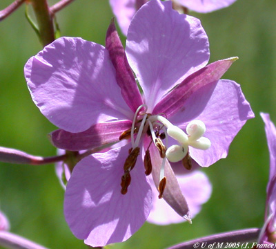
View a slide of a young flower bud of Lilium cut in cross section
Note the single compound pistil in the centre of the section. The pistil is composed of three fused carpels. Two dense staining ovule primordia are evident in each of the three loculi. Outside the pistil are six bilobed anthers, three petals and three sepals.
Having examined the prepared slide of the lily flower bud you should be able to construct a floral diagram for Lilium.
 The Anther
(Campbell 6th Ed. 787, Fig. 38.4a; 7th Ed. 774, Fig. 38.4a)
The anther is deeply bilobed. Within each lobe there are two microsporangia. At this stage of anther development there are numerous diploid microsporocytes (pollen mother cells) in the centre of each microsporangium. A single layer of columnar shaped young tapetum cells surrounds the central microsporocytes. The diploid tapetum cells transfer nutrients and certain cell wall materials to the developing pollen. As anther development proceeds, the microsporocytes divide by meiosis to produce a spherical tetrad of four microspores.
Examine a slide of Lilium anthers showing pollen tetrads
The red stained nucleus of each microspore in the tetrad should be readily visible.
The microspores separate from the tetrad, enlarge and form a thick two layered wall (exine and intine). Within the microspore (endosporic development) the nucleus divides by mitosis. A cell membrane develops around one of the nuclei. This is the elliptical shaped generative cell . The other nucleus is the tube cell nucleus. These two cells constitute the early microgametophyte (pollen grain) of Lilium.
Examine a slide of a Lilium anther containing the two cell pollen grains
Note the tube nucleus and intensely stained elliptical generative cell and nucleus.
The anther splits longitudinally (microsporangium dehiscence) and the pollen grains are released. They are transferred through the process of pollination to the stigma of the pistil (carpel). A pollen tube develops from an outgrowth of the inner (intine) wall of the pollen grain and begins to digest its way through the tissue of the stigma and style of the pistil (carpel). Unlike Pine, growth of the pollen tube in angiosperms is fairly rapid. The time interval between pollen tube initiation and the tube reaching an unfertilized ovule is quite variable, depending on the species. It is generally from a few hours to one to two days. During pollen tube growth in Lilium the generative cell divides once by mitosis to produce two sperm cells. The mature microgametophyte consists of the tube cell nucleus and two sperm.
The Ovule
(Campbell 6th Ed. 787, Fig. 38.4b; 7th Ed. 774, Fig. 38.4b)
Depending on the species, there may be from one to several hundred ovules produced in the ovary of each pistil (carpel). The ovule primordium is a small dome-shaped swelling that arises from the inner wall of the ovary. Two envelopes of tissue, the integuments, develop from the base of the primordium and completely enclose the megasporangium (nucellus). A micropyle is present at one end of the ovule. In the majority of angiosperms a single cell of the megasporangium, the megasporocyte (megaspore mother cell), enlarges and divides by meiosis to produce four haploid megaspore cells, three of which degenerate. The surviving megaspore enlarges and by means of three successive mitotic divisions, within the megaspore, gives rise to an eight nucleate megagametophyte (embryo-sac). Thin walls develop around six of the nuclei. At the micropyle end of the megagametophyte an egg cell is flanked by two synergids. At the opposite end of the megagametophyte are three antipodals. The large central region of the megagametophyte is referred to as the central cell and contains two polar nuclei.
Examine the slide of a longitudinal section of the mature ovule of Lilium on demonstration
Note the integuments, micropyle, megasporangium, funiculus, egg cell, synergids, polar nuclei and antipodals.
 The
pollen tube
grows up the micropyle
of the ovule,
through the megasporangium
and penetrates the synergid
of the megagametophyte.
The tip of the tube bursts and the sperm are released.
The
pollen tube
grows up the micropyle
of the ovule,
through the megasporangium
and penetrates the synergid
of the megagametophyte.
The tip of the tube bursts and the sperm are released.
A unique feature of the angiosperms is the process of "double fertilization". One sperm fuses with the egg to produce the zygote, the other sperm migrates to the central cell and fuses with the two polar nuclei (fusing nucleus) to produce the primary endosperm nucleus.
With double fertilization a number of processes are initiated:
|
 The Angiosperm Fruit
After fertilization the ovary component of the pistil matures into a fruit. Additional parts of the flower (e.g. the receptacle) may also contribute to the formation of the fruit (e.g. strawberry). The different types of angiosperm fruits are far too numerous and varied to be examined in any detail in this course. In general fruits may be classified as simple, aggregate or multiple ). At maturity, a given fruit may be dry or fleshy, dehiscent or indehiscent.
Whatever the type, the angiosperm fruit serves two important functions:
1. Protect the seeds during their maturation
2. Effectively disperse the mature seeds
The chief agents in seed and fruit dispersal are wind, water and animals.
![]()